
Robot-assisted sacral colpopexy
The sacral colpopexy operation is often referred to as the ‘gold standard’ operation for prolapse because of the high long-term success rates associated with this surgery. This operation was first performed in 1962 through an abdominal incision. Since 1991 this surgery has been performed using laparoscopy (keyhole surgery) and since 2004 it has been performed with robotic assistance using the da Vinci robot.
Robot-assisted sacral colpopexy is an operation that suspends the vaginal apex, using a synthetic or biological graft, from a ligament on front of the sacrum (tail bone). This provides support for the upper part of the vagina (vaginal vault). A repair inside the vagina may also be required at the same time. This operation can be performed in combination with other procedures such as surgery for urinary incontinence.
What happens during surgery?
Sacral colpopexy is an operation that suspends your vagina or uterus, using a synthetic graft or your own native tissue (fascia lata), from the front of your sacrum (tail bone). This surgery provides optimal support to the top of your vagina or uterus. A repair inside your vagina may also be required at the same time. The operation can be performed in combination with other procedures. If you prefer mesh-free surgery, tissue taken through a small incision from your thigh (fascia lata) can be used instead of mesh. Studies from Dr Carey’s own patients show that this operation successfully supports the top of the vagina or uterus in over 90% of women. This operation is usually reserved for women with severe prolapse who have already had a hysterectomy (vault prolapse).
What happens during surgery?
The surgery is performed under general anaesthetic (you are completely asleep). Dr Carey may advise you to have your operation by robotic surgery. A graft, made from your own tissue (fascia lata) or a synthetic graft, is stitched to the top of your vagina (the vaginal vault) and the other end of the graft is stitched to the front of your sacrum (tail bone). This provides very strong support for your vagina. A surgical repair of the vagina may be required, depending on the type of prolapse you have. At the end of the operation, a catheter is inserted into your bladder to drain urine and remains in place overnight. A cystoscopy (looking inside the bladder) will usually be performed at the end of the surgery to check that no damage has occurred to the bladder or ureters (the tubes running from the kidneys down to the bladder).
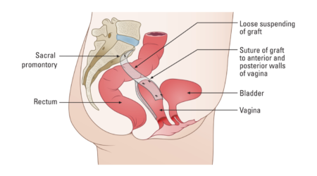
The illustration below demonstrates the vagina suspended from the sacrum with a graft (sacral colpopexy). A surgical vaginal support device (S-POP) is placed into the vagina at the end of end of surgery and is removed 21 to 28 days after surgery.
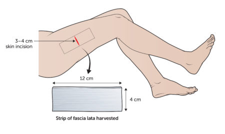
The illustration below shows that the fascia lata is taken from the outer part of your upper thigh.
 Dr Carey will be happy to answer any questions you may have and can give more specific advice. Before deciding to have surgery, you should read carefully all the information about your operation and consider obtaining a second opinion.
If you experience complications after you leave hospital, contact Dr Carey or the nursing staff on 1 West at the Epworth Freemasons Hospital for advice. In an emergency you may attend the Royal Women’s Hospital, Parkville or Epworth Hospital, Richmond emergency department or attend your closest hospital emergency department.