
Robot assisted Suture Hysteropexy and Colpopexy (Mesh Free Surgery)
Robot-assisted suture hysteropexy and suture colpopexy are both mesh free surgeries that use a patient’s own native tissue. The uterus is supported in the pelvis by the uterosacral ligaments (USL). These ligaments can tear, break or gradually become weaker which results in prolapse of the uterus. The suture hysteropexy is an operation that re-suspends the uterus from the USL by using sutures and provides support for the uterus and the upper part of the vagina. This operation is an option for women with prolapse who wish to retain their uterus.
Robot-assisted suture hysteropexy can be performed in combination with other procedures, such as surgery for urinary incontinence and vaginal prolapse (cystocele and rectocele).
Robot-assisted suture colpopexy re-suspends the vaginal apex from each USL. This operation is used for women who have had a hysterectomy or who have a hysterectomy during their prolapse surgery.
What happens during surgery?
The surgery is performed under general anaesthesia (you are completely asleep). The operation is performed using the da Vinci robotic system (key-hole surgery). The uterus or vaginal apex is suspended, using sutures, from each USL A surgical repair inside the vagina may also be required, depending on the type of prolapse you have. At the end of the operation, a catheter will be inserted into the bladder to drain urine. This will remain in place over night. A cystoscopy (looking inside the bladder with a telescope) will usually be performed at the end of the surgery to check that no damage has occurred to the bladder or ureters (the tubes running from the kidneys down to the bladder).
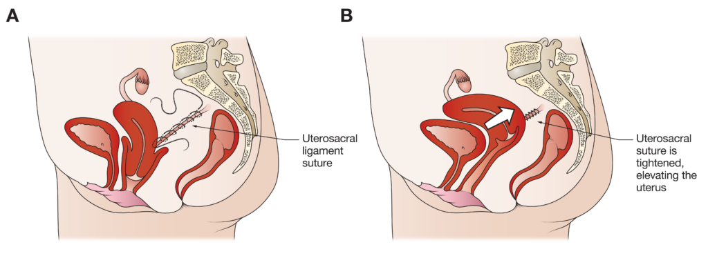
The illustrations above demonstrate a side view of the uterus suspended from each uterosacral ligament (USL) by sutures. A) Sutures are passed through each USL; B) The uterus becomes re-supported after the sutures are tied. A surgical vaginal support device (S-POP) is placed into the vagina at the end of end of surgery and is removed 21 to 28 days after surgery.
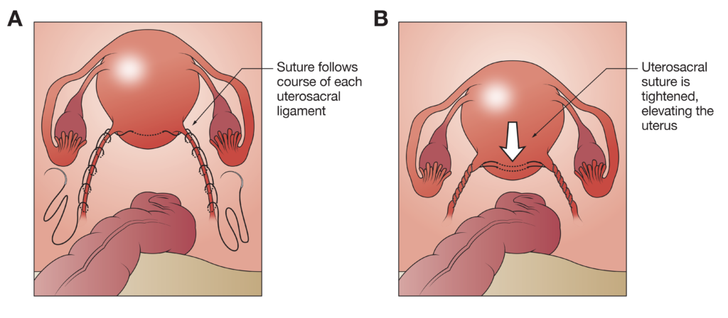
The illustrations above demonstrate a view from above the pelvis of the uterus suspended from each uterosacral ligament (USL) by sutures. A) Before the sutures are tied; B) After the sutures are tied.
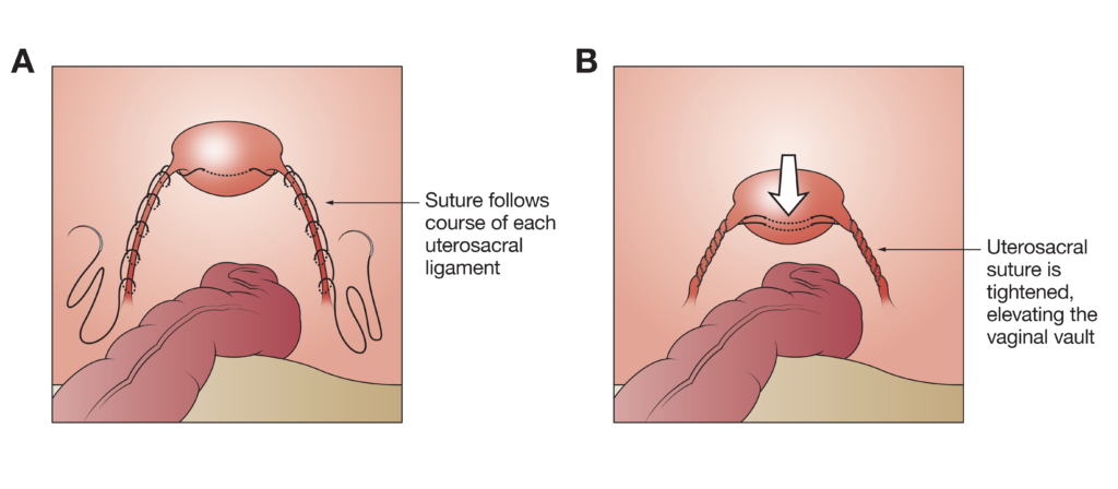
The illustrations above demonstrate a view from above the pelvis of the uterus suspended from each uterosacral ligament (USL) by sutures. A) Before the sutures are tied; B) After the sutures are tied.
 Dr Carey will be happy to answer any questions you may have and can give more specific advice. Before deciding to have surgery, you should read carefully all the information about your operation and consider obtaining a second opinion.
If you experience complications after you leave hospital, contact Dr Carey or the nursing staff on 1 West at the Epworth Freemasons Hospital for advice. In an emergency you may attend the Royal Women’s Hospital, Parkville or Epworth Hospital, Richmond emergency department or attend your closest hospital emergency department.